
Acute Shoulder Dislocation

What is a Dislocated Shoulder?

The shoulder is the most mobile joint in the body and thus susceptible to being dislocated or partially dislocated (aka subluxed). [1] The shoulder is classified as a ball and socket joint composed of three different bones: humerus, scapula and clavicle. Joints are classified as either stable joints, think knee (only moves in one direction) or a mobile joint like the shoulder or hip. Because of this mobility, stability of the joint is compromised and provided by not only the ligaments but also muscles known as the rotator cuff. These are the important dynamic restraints of the shoulder.

In the case of a shoulder dislocation, the ball (humerus) is forcefully put of out its position on its socket (glenoid labrum). This is typically associated with significant pain and an inability to use your arm until the shoulder is relocated. A shoulder subluxation is an instability event where the humerus will slide partly away from the glenoid labrum and return before a full dislocation occurs
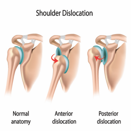
Dislocated shoulder anatomy.png

Types of Dislocations

When the shoulder is dislocated from the joint it can occur in 3 different ways dependent upon the way that the humerus moves away from the glenoid.

·      Anterior Dislocation – most common type accounting for 95-97% of cases caused by an arm being placed in a position of abduction and external rotation like making a tackle and the shoulder displaces forwards [2]

·      Posterior Dislocation – accounts of approximately 2-4% of cases and occur with the humerus displaced towards the back of the body. This can occur with a fall on an outstretched arm or elbow [3]

·      Inferior Dislocation – the rarest type where the top of the humerus is displaced downwards, typically occurs via trauma to the shoulder.

Causes

The most common cause of shoulder dislocation is trauma through force applied to the shoulder in various angles which causes the humerus to leave the glenoid labrum. These injuries commonly in contact sports such as Rugby and AFL, Eg. When making a tackle or when contact occurs contesting for a mark. 

In addition, some people who have looser ligaments throughout their body who are able to dislocate their shoulder with relative ease. The best thing for this population is to do a targeted strengthening program to reduce the risk of dislocations occurring with repetitive shoulder movements. [4]

Management of Shoulder Dislocations

Once the shoulder is relocated, there are multiple factors that will determine which direction someone may choose to proceed with treatment. However normally non-operative rehabilitation is the first line of management. An assessment with a trained Sports Physio or Specialist Sports Doctor with the appropriate imaging can help decide the best course of management in each case.

Non-Operative Management

Conservative management is the first line of treatment in most cases of first time acute shoulder dislocation. The main goals in this type of management are

·      Pain relief – reduce localised pain and reduce swelling (Rest Ice Compression Elevation - RICE)

·      Physiotherapy - Maintain and restore muscular strength and proprioceptive feedback. Regain normal range of motion once pain has settled.

·      Rehab - Increase strength to prevent further instability episodes and withstand the forces placed on the shoulder during sport and in daily life.

·      Return to full function with prevention strategies to ideally avoid a recurrent dislocation

Surgical Management

Surgical management for this injury is common in recurrent dislocators and those returning to contact sports. These shoulder reconstructive surgeries are very successful in restoring stable anatomy allowing the athlete to their chosen sport competing at the same level. A sling will be applied post-surgery for a short period of time before the required physiotherapy. Strength rehabilitation is essential in restoring the shoulder to its pre-injury status and to prevent further injury. Rehab timeframes can last from 4-9 months depending on various factors.

Written By Sports Physiotherapist Simon Wybenga

References

1.     Dodson, CC et al. Anterior glenohumeral hoint dislocations. Orthopaedic Clinical North America, 2008. 39(4): p507-18

2.     Boone, JL et al. First time anterior shoulder dislocations: has the standard changed? British Journal of Sports Medicine, 2010. 44: p355-36

3.     Farrar NG et al. An overview of shoulder instability and its management. Open Orthopaedic Journal, 2013.  7: p338-346

4.     Cutts, S et al. Anterior shoulder dislocation. Ann R Coll Surg England, 2009. 91(1): p2-7

5.     Wang, RY et al. The recognition and treatment of first-time shoulder dislocation in active individuals. JOSPT, 2009. 39(2):p118-123
Iliotibial Band Syndrome

Introduction

Running related injuries (RRI’s) are common in long distance runners with injury incidence shown to range between 17.8 (95% CI 16.7-19.1) injuries per 1000 hours of running in novice runners and 7.7 (95% CI 6.9-8.7) injuries per 1000 hours of running in recreational runners.[1] Known risk factors for RRI’s include history of previous injury and a greater training distance per week while a gradual increase in training distance per week is a protective factor. [2]

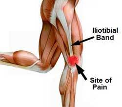
ITB Syndrome Anatomy

ITB Syndrome Anatomy

Cause

Iliotibial band syndrome (ITBS) is the second most common RRI and the most common cause of lateral knee pain in runners.[3] The aetiology of ITBS is thought to be multifactorial and remains poorly understood however several biomechanical risk factors have been proposed to contribute to the condition.[4] Traditionally, ITBS was thought to be caused by friction or excessive pressure at the distal iliotibial band (ITB) as it passes over the lateral femoral epicondyle during repetitive knee flexion and extension. [5] A more recent theory suggests that impingement at this site contributes to the condition rather than purely excessive lateral pressure.[6, 7]

Biomechanical Risk Factors

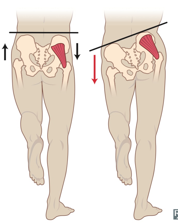
Proposed biomechanical risk factors for ITBFS include increased peak hip adduction,[8] knee internal rotation,[8-10] peak trunk ipsilateral flexion,[10, 11] and peak rearfoot pronation[12] as these factors can increase abnormal lengthening of the ITB (i.e. excess strain) and contribute to compression of the ITB at the lateral femoral condyle (i.e. excess stress).[13]

Poor Biomechanincs (Right) can contribute to ITBS

Poor Biomechanincs (Right) can contribute to ITBS

Prevention and Management

Preventative measures for ITBFS should include an appropriate training plan that includes gradual increases in running distance and appropriate load management strategies including programmed rest days and de-load phases.[2]

Management

The acute management of ITBS should focus on anti-inflammatory modalities, relative rest, soft tissue release and addressing biomechanical factors known to contribute to ITBS.[13] As irritability settles running can be gradually re introduced in line with a periodised running plan. Running retraining incorporating running with an increased cadence has been shown to increase the gluteal muscle recruitment during foot strike and mid stance of the running cycle [14] and may help to decrease load at the ITB insertion in patients with ITBS.


There is debate as to whether it is possible to modify running mechanics with strengthening exercises alone however positive results have been demonstrated in studies that focus on isometric and eccentric strength of the glute muscles in running specific positions[15] and include visual feedback with cueing.[16] The hip abductor muscles are known to contribute to peak hip adduction, knee internal rotation, ipsilateral trunk flexion[17] and have been shown to be related to other lower limb RRI’s.[14] Two gluteal muscles that control frontal and transverse plane hip and pelvic motion during running include the glute max and glute med.[14] Glute max EMG is shown to be highest in the step up and single leg squat exercise while the glute med EMG is shown to be highest in the side bridge with hip abduction exercise [18] (See Pictures Below). Despite a lack of prospective research it is plausible to include gluteal strengthening 2-3 x per week in the management of ITBS given the gluteal muscles’ relationship to biomechanical factors known to be associated with the condition.


While foot and ankle orthosis have been proposed as a tool to maintain a neutral foot position and control tibial internal rotation during running[19] to date, no prospective studies have investigated the use of orthosis for patients with ITBS.

Single Leg Squat

Single Leg Squat

Side Bridge with Hip Abduction

Side Bridge with Hip Abduction

Conclusion

Based off the available evidence the best management for ITBS includes appropriate management in the acute stage to settle irritability followed by gluteal strengthening with an emphasis on isometric and eccentric frontal and transverse plane movements and physiotherapist guided running retraining.

Take Away Points

·      ITBS is the second most common RRI and the most common cause of lateral knee pain in runners.

·      ITBS is thought to be caused by either excessive lateral pressure or impingement as the ITB runs over the lateral femoral epicondyle during knee flexion and extension.

·      Gluteal weakness is proposed as a modifiable contributing factor in ITBS.

·      Gluteal strengthening should focus on isometric and eccentric exercises in running specific positions and should be carried out with appropriate cueing and visual feedback.

·      Running retraining including running with an increased cadence has been proposed to increase gluteal muscle activation during running.

Written by Cameron Dyer – Physiotherapist and Athlete Rehab Specialist


References

1.         Videbæk, S., et al., Incidence of Running-Related Injuries Per 1000 h of running in Different Types of Runners: A Systematic Review and Meta-Analysis. Sports Medicine, 2015. 45(7): p. 1017-1026.

2.         van Gent, R.N., et al., Incidence and determinants of lower extremity running injuries in long distance runners: a systematic review. British Journal of Sports Medicine, 2007. 41(8): p. 469-480.

3.         Taunton, J.E., et al., A retrospective case-control analysis of 2002 running injuries. British Journal of Sports Medicine, 2002. 36(2): p. 95.

4.         Beers, A., et al., Effects of Multi-modal Physiotherapy, Including Hip Abductor Strengthening, in Patients with Iliotibial Band Friction Syndrome. Physiotherapy Canada. Physiotherapie Canada, 2008. 60(2): p. 180-188.

5.         Orchard, J.W., et al., Biomechanics of Iliotibial Band Friction Syndrome in Runners. The American Journal of Sports Medicine, 1996. 24(3): p. 375-379.

6.         Hamill, J., et al., A prospective study of iliotibial band strain in runners. Clinical Biomechanics, 2008. 23(8): p. 1018-1025.

7.         Fairclough, J., et al., Is iliotibial band syndrome really a friction syndrome? Journal of Science and Medicine in Sport, 2007. 10(2): p. 74-76.

8.         Noehren, B., I. Davis, and J. Hamill, ASB clinical biomechanics award winner 2006 prospective study of the biomechanical factors associated with iliotibial band syndrome. Clinical biomechanics (Bristol, Avon), 2007. 22(9): p. 951.

9.         Ferber, R., et al., Competitive female runners with a history of iliotibial band syndrome demonstrate atypical hip and knee kinematics. The Journal of orthopaedic and sports physical therapy, 2010. 40(2): p. 52.

10.       Foch, E., et al., Associations between iliotibial band injury status and running biomechanics in women. Gait & Posture, 2015. 41(2): p. 706-710.

11.       Foch, E. and C.E. Milner, The influence of iliotibial band syndrome history on running biomechanics examined via principal components analysis. Journal of Biomechanics, 2014. 47(1): p. 81-86.

12.       Messier, P.S., et al., Etiology of iliotibial band friction syndrome in distance runners. Medicine & Science in Sports & Exercise, 1995. 27(7): p. 951-960.

13.       Baker, R.L. and M. Fredericson, Iliotibial Band Syndrome in Runners: Biomechanical Implications and Exercise Interventions: Biomechanical Implications and Exercise Interventions. Physical Medicine & Rehabilitation Clinics of North America, 2016. 27(1): p. 53-77.

14.       Semciw, A., R. Neate, and T. Pizzari, Running related gluteus medius function in health and injury: A systematic review with meta-analysis. Journal of Electromyography and Kinesiology, 2016. 30: p. 98-110.

15.       Fredericson, L.M., et al., Hip Abductor Weakness in Distance Runners with Iliotibial Band Syndrome. Clinical Journal of Sport Medicine, 2000. 10(3): p. 169-175.

16.       Barrios, J.A., K.M. Crossley, and I.S. Davis, Gait retraining to reduce the knee adduction moment through real-time visual feedback of dynamic knee alignment. Journal of Biomechanics, 2010. 43(11): p. 2208-2213.

17.       Mucha, M.D., et al., Hip abductor strength and lower extremity running related injury in distance runners: A systematic review. Journal of Science and Medicine in Sport, 2017. 20(4): p. 349-355.

18.       Macadam, P., J. Cronin, and B. Contreras, An examination of the gluteal muscle activity associated with dynamic hip abduction and hip external rotation exercises. A systematic review. International journal of sports physical therapy, 2015. 10(5): p. 573.

19.Aderem, J. and Q.A. Louw, Biomechanical risk factors associated with iliotibial band syndrome in runners: A systematic review Rehabilitation, physical therapy and occupational health. BMC Musculoskeletal Disorders, 2015. 16(1): p. <xocs:firstpage xmlns:xocs=""/>.

Achilles Tendon Pain And Isometrics

Tendons

Tendon injuries and pain are common amongst athletes of various sports and ages. Tendons attach muscle to bones and allow muscles to transmit force across our joints which allow us to move, walk, run and jump!

Tendons most commonly become injured when they undergo a period of loading above and beyond what they are used to. We call these injuries Tendinopathies and they have been shown to have a continuum of dysfunction ranging from reactive to degenerative, or more commonly described as acute or chronic (Cook and Purdam 2008). Due to this variability of tendinopathy injuries, management for tendon injuries can be difficult and requires accurate assessment and targeted individualised interventions.

Isometrics

Current research in patella tendons has shown isometric exercise has a place in athletes with pain as it has an analgesic effect (Rio et al 2017). The findings of these studies in patella tendons have been applied for tendons throughout the body in both professional sports and in private practice with the general population. The proposed cause of pain relief with isometrics in these studies is a reduction in pain related muscle inhibition leading to increase in strength and changes to the central nervous system which dampen pain. (Rio et al 2015)

More recently it has been proposed that this widespread adoption of isometrics may not be applicable to tendons throughout the body in areas such as the Achilles tendon, lateral epicondylalgia (tennis elbow) or hamstring tendons (Coombes et al 2016).

Achilles Tendon Anatomy

Achilles Tendon Anatomy

Muscles That Attach to the Achllies - Gastrocnemius Vs Soleus

There are 3 muscles that attached to the Achilles Tendon. The soleus and the gastrocs are the largest and have the most significant contribution to athletes who run, jump and change direction. The gastroc’s crosses the knee and is a more active in ankle movements when the knee is straight, and the soleus is more active when the knee is bent. The reality is that both muscles are quite different in their location and function, but they both need the be strong and work together to support the Achilles Tendon during sport.

Calf Muscle Anatomy

Calf Muscle Anatomy

People with tendon injuries and tendon pain have been found to have weaker calf strength in both the injured and the corresponding limb muscles than other people without tendon pain (O’Neill 2018). Essentially if an athlete does not have the adequate strength in their muscle, their tendon will be required to take on more load. As a result we often see them fail and become painful as they attempt to cope with the excessive loading that the tendon is unaccustomed to. So STRENGTH is the key! But which type???

Other Exercise Therapy

Isotonic (Or through range strength training) strength training, which includes both concentric and eccentric components, has recently been suggested as a more appropriate way to manage tendon injuries other than the patella tendon (O’Neill et 2018).  This type of strength training needs to be heavy at around 70-80% of 8RM (repetitions maximum), and must be appropriate to the type and stage of tendon injury as detailed above. In Achilles Tendinopathies the most appropriate exercises are loaded calf raise in seated and standing positions (see pictures below). These should be progressed to single leg variations with load as symptoms and strength allows. Both of these exercises target the major calf muscles of the gastrocnemius and soleus in differing amounts.

*NOTE: These aren’t intended to be pain relieving and need to be prescribed around the athletes training appropriately.

Loaded Standing Single Leg Calf Raise

Loaded Standing Single Leg Calf Raise

Loaded Seated Calf Raise

Loaded Seated Calf Raise

To Isometric Or Not

The type of strength stimulus we put through our tendon and adjoining muscles is extremely important when dealing with injury and needs to be case specific. As mentioned Isometric strength training has been successfully used for tendinopathies in certain areas of the body, and as a result adapted to other areas throughout the body.

However, they are not the only form of resistance training we should expose to an injured tendon, as strength gain’s through ROM are important and functional to the sports that many athletes play. As for the Achilles the focus should be on improving strength in the calf complex relative to the athlete’s sport. This may be a combination of loaded strength rehab and higher velocity power strength work. This ALWAYS needs to be specific to the demands of the athlete’s sport and goals.

Isometric exercises still have an important role in tendon pathology!! But they aren’t the only type of strength to take into consideration when you have any type of tendon pain.

For other general information regarding tendons and in particular patella tendons refer to our previous article published by Luke Heath on our website “Jumpers Knee: Patella Tendiopathy – Diagnosis and Management”.

For any further questions or to book an appointment contact us online or via phone.

Written By Chris Bailey - Titled Sports Physiotherapist and Rehab Specialist


References

·      Cook J, Purdam C. Is Tendon Pathology a Continuum? A Pathology Model to Explain the Clinical Presentation of Load-Induced Tendinopathy. British Journal of Sports Medicine. 2009

·      Coombes B, Tucker K, Vincenzino B. Achilles and Patella Tendinopathy Display Opposite Changes on Elastic Properties: A Shear Wave Elastograpy Study. Scandavian Journal of Medicine and Science in Sports. 2018 

·      O’Neill S, Radia J, Bird K, Rathleff M, Bandholm T, Jorgensen M, Thorborg K. Acute Sensory and Motor Response to 45-s Heavy Isometric Holds for the Plantar Flexors in Patients with Achilles Tendinopathy. Journal of Knee Surgey, Sports Tramatology and Arthroscopy. 2018

·      O’Neill S. PhysioEdge podcast Episode 82. 2018

·      O’Neill S, Watson P, Barry, S. Why are Eccentric Exercises Effective for Achilles Tendinopathy? The International Journal of Sports Physical Therapy. 2015

·      Rio E, Kidgell D, Purdam C, Gaida J, Mosely L, Pearce A, Cook J. Isometric Exercise Induces Analgesia and Reduces Inhibition in Patella Tendinopathy. British Jounal of Sports Medicine. 2015

·      Rio E, Van Ark M, Docking S, Mosley L, Kidgell D, Akker-Scheek I, Zwerver J, Cook J. Isometric Contractions are More Analgesic than Isotonic Contractions for Patella Tendon Pain: An In-Season Randomised Clinical Trail. Clinical Journal of Sports Medicine. 2017

Strength Training for Endurance Sports Performance

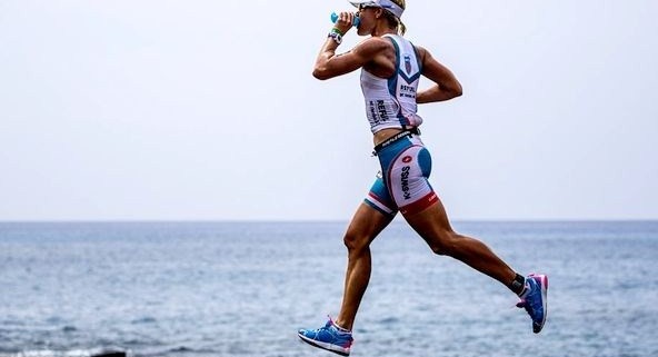
strive4fitnessuk.com Mirinda Carfrae.jpg

Strength training remains an under-utilised tool to improve performance in endurance sports despite recent evidence showing beneficial outcomes (Blagrove, Howatson, & Hayes, 2018). Concerns about slowing down, gaining muscle mass and the perceived opportunity cost of spending time performing strength training rather than doing endurance training prevent many endurance sports athletes from the benefits of strength training.

Why is this the case?

Before we discuss strength training for endurance athletes there are three primary physiological determinants of endurance sports performance that we need to acknowledge. The combination of these three factors form the primary physiological and biomechanical determinants of endurance sport outcome.

·       VO2 max – the maximal amount of oxygen that can be consumed during aerobic exercise. This can be viewed as the overall CAPACITY of the body to use oxygen to perform work.

·       Blood lactate markers – when our muscles use oxygen to create energy a waste product called lactic acid is formed. The blood lactate threshold refers to the maximal amount of oxygen that can be consumed before lactic acid accumulates and causes a reduction in work capacity.  Blood lactate markers can be viewed as the COST of muscle metabolism.

·       Economy – reflects the oxygen or energy cost of sustaining a given sub-maximal running velocity. This is a measure of how efficiently you use oxygen at submaximal levels and is determined predominantly by neuromuscular factors. Movement economy can be thought of as the overall EFFICIENCY of the body to generate energy.

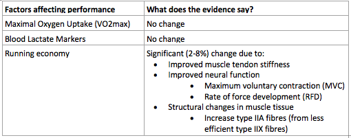
A recent systematic review by Blagrove et al (2018) (Blagrove et al., 2018) reviewed 24 studies of 469 trained (>6 months) middle and long distance runners to assess the effect of strength training on the above three physiological determinants of performance. The review only included studies that had a control group of endurance only training and included strength training interventions using either heavy resistance training, explosive resistance training or plyometric training between 1 and 4 x per week.

The results of the review are included in a table 1.

Factors affecting performance

Table 1. Results from Systematic Review of Blagrove et al (2018)

Table 1. Results from Systematic Review of Blagrove et al (2018)

The review also found that while strength training did NOT improve VO2 max or blood lactate levels, they were not negatively affected. In addition, the athletes that performed strength training achieved a higher terminal velocity in a maximal aerobic running test i.e. they improved their running velocity significantly more than matched controls that only did endurance training.

Improvements in running economy, measured as the oxygen or energy cost of running at a given speed, hold the biggest key for endurance athletes to improve their performance. Running economy was improved by three main mechanisms:

1.     Improved muscle tendon stiffness

a.     Muscle tendon stiffness refers to the bodies ability to utilise the stretch shortening cycle to generate force. Put simply, rather than the muscles having to generate all the force to propel the body forward each step, the muscle tendon unit can store elastic energy from the previous step and ‘release’ it into the following step. Think of an elite runner and how they seem to effortlessly bounce along the track verses a beginner that plods along. Who is running more efficiently?

2.     Improved neural function

a.     Strength can improve by two primary ways:

                                               i.     Greater coordination of the muscle unit

                                              ii.     Larger cross-sectional area (size) of the muscle fiber

b.     Strength training in endurance athletes works by increasing the maximum voluntary contraction and the rate of force development i.e. better coordination between the nerve signal and the muscle unit

3.     Structural changes in muscle tissue

a.     There are different types of skeletal muscle fibers that each have specific qualities. Strength training converts the large fast twitch highly fatigable (type IIX) fibers to the smaller fast twitch more efficient (type IIA) fibers.

Figure 2. Proposed mechanisms by which short term and long term endurance performance can be improved from the addition of strength training to the ongoing endurance training plan. (Aagaard &amp; Andersen, 2010)

Figure 2. Proposed mechanisms by which short term and long term endurance performance can be improved from the addition of strength training to the ongoing endurance training plan. (Aagaard & Andersen, 2010)


Interestingly, including strength training did not result in an increase in muscle mass despite participants getting significantly stronger. This is because performing a high volume of endurance training stunts the intracellular hypertrophy response normally associated with resistance training (Wilson et al., 2012) (Nader, 2006). This suggests that strength training in endurance athletes improves strength via neural changes i.e. greater coordination of muscle firing and faster rate of contraction rather than by increasing the size of the muscle.

Besides the performance benefits, strength training is also a key component of most injury prevention programs. Runners are known to have a high overall musculoskeletal running related injury (MRRI) incidence of 19.4-79.3%. (van Gent et al., 2007) The most common MRRI is medial tibial stress syndrome which is known to have several strength related risk factors i.e. low calf power and poor glute medius strength (Galbraith & Lavallee, 2009). In this way, strength training not only helps to improve performance but can help in building resilience to common running related injuries.

In summary - strength training can significantly improve running economy, running velocity and reduce time lost to common running related injuries without causing an increase in weight, muscle mass or negatively affecting VO2 max or blood lactate markers.

Which leaves me with the question…


Screen Shot 2019-02-14 at 8.58.10 am.png


When integrating strength training into an endurance training plan, athletes should look to work with an experienced coach that understands your injury history, training history, goals and has experience writing strength programs for running based athletes.

Many endurance sports athletes have little to no experience with strength training and for this reason the strength training program must be planned, periodised, supervised and progressively overloaded with detail and care.

For more information about strength training for endurance sports athlete’s feel free to reach out.


Written by Cameron Dyer – Physiotherapist and Athlete Rehab Specialist


References:

Aagaard, P., & Andersen, J. L. (2010). Effects of strength training on endurance capacity in top-level endurance athletes. In (Vol. 20 Suppl 2, pp. 39).

Blagrove, R., Howatson, G., & Hayes, P. (2018). Effects of Strength Training on the Physiological Determinants of Middle- and Long-Distance Running Performance: A Systematic Review. Sports Medicine, 48(5), 1117-1149. doi:10.1007/s40279-017-0835-7

Galbraith, R., & Lavallee, M. (2009). Medial tibial stress syndrome: conservative treatment options. Current Reviews in Musculoskeletal Medicine, 2(3), 127-133. doi:10.1007/s12178-009-9055-6

Nader, A. G. (2006). Concurrent Strength and Endurance Training: From Molecules to Man. Medicine & Science in Sports & Exercise, 38(11), 1965-1970. doi:10.1249/01.mss.0000233795.39282.33

van Gent, R. N., Siem, D., van Middelkoop, M., van Os, A. G., Bierma-Zeinstra, S. M. A., Koes, B. W., & Taunton, J. E. (2007). Incidence and determinants of lower extremity running injuries in long distance runners: a systematic review. British Journal of Sports Medicine, 41(8), 469-480. doi:10.1136/bjsm.2006.033548

Wilson, M. J., Marin, J. P., Rhea, R. M., Wilson, M. C. S., Loenneke, P. J., & Anderson, C. J. (2012). Concurrent Training: A Meta-Analysis Examining Interference of Aerobic and Resistance Exercises. Journal of Strength and Conditioning Research, 26(8), 2293-2307. doi:10.1519/JSC.0b013e31823a3e2d


Plantar Fasciopathy

Background

Plantar fasciopathy (previously known as plantar fasciitis) is the most common cause of heel pain in adults and affects up to 8% of running-based athletes. (Taunton et al., 2002) Symptoms of plantar fasciopathy include pain at the base of the heel that is worst in the morning or when performing sporting activity, particularly when warming up. (Thing, 2012) Plantar fasciopathy usually results from an acute overload of the plantar fascia i.e. when commencing a new sport or a chronic overload related to lifestyle and exercise factors. Risk factors for plantar fasciopathy include reduced ankle dorsi flexion, BMI > 30kg/m2 and work-related weight-bearing.(Riddle, Pulisic, Pidcoe, & Johnson, 2003)

Recent evidence suggests that the condition should be referred to as fasciopathy rather than fasciitis as the tissue pathology more closely resembles that of tendinosis. (Drew, Smith, Littlewood, & Sturrock, 2014; Lemont, Ammirati, & Usen, 2003)

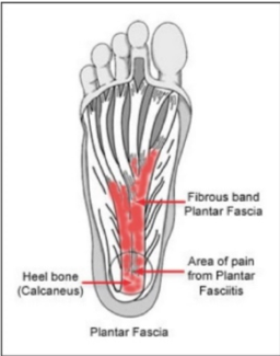
Plantar Fascia Anatomy

Plantar Fascia Anatomy

Assessment and Diagnosis

Plantar fasciopathy is generally diagnosed clinically however ultrasound can assist if clinical diagnosis is uncertain. Ultrasound can be used to measure the thickness of the fascia however ultrasound findings have not been shown to change despite improvements in outcome. (Rathleff et al., 2015) In some case, patients with plantar fasciopathy present to physiotherapy with an X-Ray that may show a bone spur at the base of the heel. Interestingly, this has not been shown to be related to pain or outcome therefore is generally not of use in guiding management.(Lu, Gu, & Zhu, 1996)

Treatment and Rehabilitation

Treatment of plantar fasciopathy has traditionally consisted of stretching, gel heel inserts and injections (Rathleff & Thorborg, 2015) however recent advances in understanding of the pathology has led to increased focus on exercise and load management as treatment options.

A recent study by Rathleff et al, (2015) showed that a program of slow high-load strength training outperformed a stretching program over 3 months. The strength program consisted of progressively overloading a single leg calf raise performed in toe flexion (Image 1/Image 2) and progressing every second week. It is a high load slow tempo strength program. Stretching consisted of 10 x 10 sec stretches performed 3 x per day.

Start position

Start position

End position

End position

The rationale behind the slow high load strength training program is that the program provides a progressive overload of the plantar fascia that promotes collagen synthesis to help normalise tendon structure and improve tissue capacity.(Rathleff et al., 2015)

While other management options do exist and are called in some cases of plantar fasciopathy the mainstay of effective management should be an appropriate load management and strengthening program administered and reviewed regularly by your physiotherapist.

Patient Tips

·       Increase the load on the plantar fascia by filling a back pack with books as you get stronger.

·       Perform the single leg calf raise with a rolled towel under your toe to utilise the ‘windlass mechanism’ whereby flexing the toes causes tensioning of the plantar fascia prior to loading.

·       Manage your activities around your pain threshold i.e. pain > 4/10 during or after loading suggests you have overloaded the tissue. For best results, discuss with your sports physiotherapist about an individualised load management plan.

Clinical Tip

·       When treating athletes or patients with plantar fasciopathy treatment outcomes are best measured using the Foot Function Index (FFI). The FFI consists of 23 items divided into three subscales that quantify the impact of foot pathology on pain, disability and activity limitations. Scores range from 0-230 with a minimal important change of 7 points for the total scale.

Written By Cameron Dyer

References

Drew, B. T., Smith, T. O., Littlewood, C., & Sturrock, B. (2014). Do structural changes (eg, collagen/matrix) explain the response to therapeutic exercises in tendinopathy: a systematic review. British Journal of Sports Medicine, 48(12), 966. doi:10.1136/bjsports-2012-091285

Lemont, H., Ammirati, K. M., & Usen, N. (2003). Plantar fasciitis: a degenerative process (fasciosis) without inflammation. Journal of the American Podiatric Medical Association, 93(3), 234. doi:10.7547/87507315-93-3-234

Lu, H., Gu, G., & Zhu, S. (1996). Heel pain and calcaneal spurs. Zhonghua wai ke za zhi [Chinese journal of surgery], 34(5), 294.

Rathleff, M. S., Molgaard, C. M., Fredberg, U., Kaalund, S., Andersen, K. B., Jensen, T. T., . . . Olesen, J. L. (2015). High-load strength training improves outcome in patients with plantar fasciitis: A randomized controlled trial with 12-month follow-up.(Report). 25(3), e292.

Rathleff, M. S., & Thorborg, K. (2015). ‘Load me up, Scotty’: mechanotherapy for plantar fasciopathy (formerly known as plantar fasciitis). British Journal of Sports Medicine, 49(10), 638. doi:10.1136/bjsports-2014-094562

Riddle, L. D., Pulisic, E. M., Pidcoe, E. P., & Johnson, E. R. (2003). Risk Factors for Plantar Fasciitis: A Matched Case-Control Study. The Journal of Bone & Joint Surgery, 85(5), 872-877. doi:10.2106/00004623-200305000-00015

Taunton, J. E., Ryan, M. B., Clement, D. B., McKenzie, D. C., Lloyd-Smith, D. R., & Zumbo, B. D. (2002). A retrospective case-control analysis of 2002 running injuries. British Journal of Sports Medicine, 36(2), 95. doi:10.1136/bjsm.36.2.95

Thing, J. (2012). Diagnosis and management of plantar fasciitis in primary care. British Journal of General Practice, 62(601), 443-444. doi:10.3399/bjgp12X653769

ACL Rehab & Preventing Re-Injury

ANATOMY & BACKGROUND

Anterior Cruciate Ligament (ACL) injuries are very common in Australian sports and lead to a significant amount of time on the sidelines for the athlete. The majority of people who wish to return to their sport opt for surgical management to replace their ruptured ACL. There is still a high percentage of these injuries that undergo reconstructive surgery that re-rupture with return to sport, up to as high as 30% (Grindum et al, 2016).

The majority of ACL injuries occur as non-contact injuries. Most commonly when an athlete is sidestepping, landing from a jump, decelerating or changing direction (Waters, 2012).

Anterior Cruciate Ligament Anatomy

Anterior Cruciate Ligament Anatomy

ACL REHABILITATION

A safe return to sport while reducing the chance or re-injury injury rates requires a comprehensive and structured rehab program designed and implemented by a rehab specialist. It is important to address the initial cause that contributed to the ACL injury in the first place, whether that is strength, cutting mechanics or poor landing or decelerating mechanics these need to be worked on frequently before returning to your chosen sport. Objective lower limb and knee assessments such as Pitch Ready can significantly help guide this decision-making process.

Image Credit: www.pitch-ready.com/

Image Credit: www.pitch-ready.com/

TIME & STRENGTH

Two crucial factors for return to sport following ACL reconstructive surgery are TIME post-op and regaining normal STRENGTH in your lower limb. Good research has showed returning to sport after 9 months post-op can significantly reduce the re-injury rate.

Quadricep strength has also been shown to be a strong predictor for safer return to sport. Both TIME and quadriceps STRENGTH together combine to reduce ACL re-injury by over 80% (Grindem et al, 2016).

Most importantly returning to sport following ACL surgery should be made on a case by case basis following guidance from your sports physiotherapist and ultimately your surgeon.

Lower Limb Strength Rehab

Lower Limb Strength Rehab

OTHER FACTORS

There are many other factors that need to be covered in your rehabilitation from an ACL operation beyond the time you take to return to sport and your quads strength. Other variables including your jump and land mechanics, cutting technique, lumbopelvic and hamstring strength. These as well as many others need to be taken into account when decisions around returning to COD sports are made. Structured assessment of your movement, strength and power are also extremely important to help guide your safe return to sport.


Written By Chris Bailey

References

  • Grindum H, Snyder-Mackler L, Moksnes H, Engebresten L & Risberg M. Simple decision rules can reduce reinjury risk by 84% after ACL reconstruction: the Delaware-Oslo ACL cohort study. British Journal of Sports Medicine (2016) 50. 804-808.

  • McGrath T, Waddington G, Scarvell J, Ball N, Creer R, Woods K, Smith D & Adams R. An ecological study of Anterior Cruciate Ligament Reconstruction, Part 1. The Orthopaedic Journal of Sports Medicine (2016) 4. 1-7.

  • McGrath T, Waddington G, Scarvell J, Ball N, Creer R, Woods K, Smith D & Adams R. An ecological study of Anterior Cruciate Ligament Reconstruction, Part 2. The Orthopaedic Journal of Sports Medicine (2017) 5. 1-9.

  • Pitch Ready Webpage. http://www.pitch-ready.com/

Hamstring Injuries and Rehabilitation

ANATOMY AND BACKGROUND

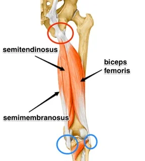
The hamstring muscle group consists of 3 major muscles in the posterior thigh, that play crucial functions in activities involving running and sprinting, and also kicking.

Soft tissue hamstring injuries make up a large proportion of time lost to injury in field based sports (Orchard & Seward, 2010), and traditionally have a very high recurrence rate of up to 30%. The most commonly injured of the hamstring muscles is the lateral biceps femoris muscle.

Hamstring Musculature

Hamstring Musculature

CONSEQUENCES OF HAMSTRING MUSCLE INJURIES

Current research shows once you have injured your hamstring muscle, you are at an increased risk of re-injuring your hamstring in the future.

Due to a combination of factors including chronic muscle inhibition and wasting, shortening of your muscle fibres, potential neural changes in the way your muscles are firing, and a drop off in eccentric muscle strength, your chances of further hamstring muscle injury is increased (Opar et al, 2018). This re-injury may also happen at a load lower than that which caused your initial injury.

Thigh Muscles Cross-Section

Thigh Muscles Cross-Section

ECCENTRIC TRAINING AND REHAB

Strength training and rehab running appropriate to your stage of recovery are integral components of getting you back to your chosen sport as quickly and safely as possible, while minimising your risk of injury recurrence (Bourne et al, 2017). Physiotherapy rehab programs targeting lumbopelvic strength as well as hamstring strength is heavily supported by the research (Heiderscheit et al 2010).

Eccentric strength in particular has been shown to improve hamstring muscles strength through greater ranges of motion, and also to increase the hamstring muscles overall length which in turn reduces the likelihood of hamstring muscle strain.

The Nordic Hamstring Exercise and Hip Extension exercises are great rehab exercises to achieve these goals. See Pictures below.

Nordic Hamstring Exercise

Nordic Hamstring Exercise

Hip Extension Exercise

Hip Extension Exercise

REHABILITATION AND PAIN

Traditionally hamstring and posterior thigh pain has been avoided during the rehab phases following hamstring injury. It has now been shown that a tolerable amount of pain during strength rehab may not only be ok, but help get the injured muscle stronger quicker, and allow for a safer and faster return to running, sprinting and your sport (Hickey 2017).

This rehab should however be done under the guidance of a highly trained Sports Physio to teach you the correct exercises and technique. And also to help guide you through which pain is acceptable to push through, to get you back to your sport stronger.

Written By Chris Bailey


Reference List

·      Bourne M, Duhig S, Timmins R, Williams M, Opar D, Najjar A, Kerr G, Shield A. Impact of the Nordic hamstring and hip extension exercises on hamstring architecture and morphology: implications for injury prevention.British Journal of Sports Medicine. 2017

·      Heiderscheit BC, Sherry MA, Silder A, Chumanov ES, Thelen DG. Hamstring strain injuries: recommendations for diagnosis, rehabilitation, and injury prevention. Journal of Orthopaedics and Sports Physical Therapy. 2010

·      Hickey J. PhysioEdge podcast Episode 72. 2017

·      Orchard JW, Seward H. Injury Report 2009: Australian Football League. Sport Health. 2010

·       Opar D, Williams M, Timmins R, Dear N, Shield A. Knee flexor strength and bicep femoris electromyographical activity is lower in previously strained hamstrings. Journal of Electromyography and Kinesiology. 2013

Jumpers Knee: Patellar Tendinopathy - Diagnosis & Management

Patellar Tendinopathy (PT) or Jumpers Knee is a common injury in jumping sports that occurs when loads are very high or suddenly increase. It affects an athlete’s performance ability to jump, land, change direction and run. It can lead to a reduction in tolerance of training and competition load and eventually a decline in performance, missing training and competition.

The aim of this article is to guide you through understanding why you have a PT, and how best to manage your grumbling patella tendon.